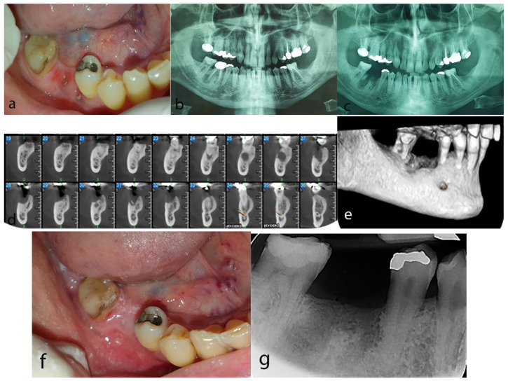
Figure 3.
Case 3: (a) Exposed necrotic bone at the site of the extraction of the first right mandibular molar. (b) Panoramic radiograph before the extraction revealing radiolucent lesions around the apices of the right first mandibular molar. (c) Post-extraction panoramic radiograph showing phantom socket at the extraction site. (d,e) CBCT revealing a diffuse hypodense lesion containing a hyperdense area at the post-extraction site of the right mandible (d) cross sectional views, (e) 3D reconstruction). (f) Complete mucosal coverage at the post-extraction site after 4 months of conservative treatment. (g) Periapical radiograph demonstrating normal bone remodeling process after 4 months of conservative treatment.

