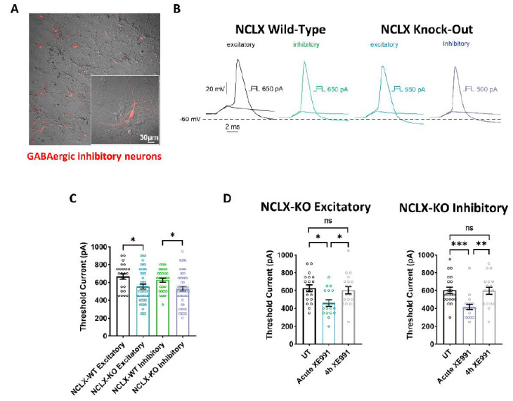
Figure 4.
Intrinsic excitability properties of hippocampal neurons derived from NCLX-WT and NCLX-KO mice. (A) Cultured hippocampal neurons infected with recombinant AAV vector driving the expression of the fluorescent protein mCherry, under the control of the specific GABAergic mDlx enhancer. GABAergic inhibitory neurons expressing mCherry are seen as red. (B) Representative traces of solitary spike discharge evoked by 2 ms step injection of depolarizing currents with increments of 50 pA in NCLX-WT excitatory neurons (black), NCLX-WT inhibitory neurons (green), NCLX-KO excitatory neurons (blue), and NCLX-KO inhibitory neurons (purple). (C) Threshold current for spike firing of NCLX-KO compared to NCLX-WT excitatory and inhibitory neurons (one-way ANOVA, F(3,150) = 4.793; Bonferroni’s multiple-comparisons test; NCLX-WT excitatory vs. NCLX-KO excitatory: *p = 0.0176; NCLX-WT inhibitory vs. NCLX-KO inhibitory: *p = 0.0268; n > 3). (D) Threshold currents of untreated NCLX-KO excitatory (left) and NCLX-KO inhibitory (right) neurons, compared to neurons acutely exposed to 10 μM XE991 and after 4 h chronic exposure (excitatory: one-way ANOVA, F(2,52) = 5.251; Tukey’s multiple-comparisons test; UT vs. acute XE991: *p = 0.0108; Acute XE991 vs. 4 h XE991: *p = 0.0325; UT vs. 4 h XE991: ns; Inhibitory: one-way ANOVA, F(2,56) = 9.277; Tukey’s multiple-comparisons test; UT vs. acute XE991: ***p = 0.0010; Acute XE991 vs. 4 h XE991: **p = 0.0018; UT vs. 4 h XE991: ns; n > 3). ns—not significant. Error bars denote SEM.

