Figure 5.
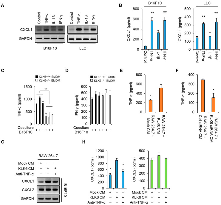
KLK6 promoted CXCL1 production by stimulating TNF-α secretion by macrophages. (A,B) B16F10 and LLC cells were treated with TNF-α (100 ng/mL), IL-1β (100 ng/mL), or IFN-γ (100 ng/mL) for 24 h. (A) The mRNA levels of CXCL1 in the indicated cytokine-treated B16F10 and LLC cells were analyzed by RT-PCR. (B) The protein levels of CXCL1 in the supernatants of the indicated cytokine-treated B16F10 and LLC cells were analyzed by ELISA. The levels of TNF-α (C) and IFN-γ (D) in the supernatants of WT or KLK6−/− BMDMs were analyzed by ELISA. (E) The secreted TNF-α levels in the supernatants of the mock- or pCMV3-KLK6-transfected RAW 264.7 cells were measured by ELISA. (F) The secreted TNF-α levels in the supernatants of the control siRNA- or KLK6 siRNA-transfected RAW 264.7 cells were measured by ELISA. (G,H) B16F10 cells were treated with the CMs of RAW 264.7 cells that were the mock vector or KLK6-expressing vector in the presence or absence of the TNF-α neutralizing antibody (2 µg/mL) for 24 h. (G) The mRNA levels of CXCL1 and CXCL2 in the indicated CM-treated B16F10 cells were analyzed by RT-PCR. (H) The protein levels of CXCL1 and CXCL2 in the supernatants of the indicated CM-treated B16F10 cells were analyzed by ELISA. Statistical significance was determined by the Student’s t-test. * p < 0.05 and ** p < 0.01. Data are representative of three experiments.

