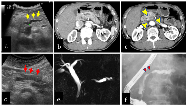
Figure 2.
Two cases of pancreatic cancer found by medical checkup by abdominal ultrasonography. (a–c): Sixty-five-year-old man had a dilatation of the main pancreatic duct (MPD) in the body (yellow arrows) and underwent enhanced computed tomography and pancreatic head cancer was found (yellow arrowheads). He had surgery and a diagnosis of stage 2b (UICC 8th) and was alive without relapse 2621 days after surgery. (d–f): An eighty-year-old women had a dilatation of MPD in the pancreatic body (red arrows) and underwent magnetic resonance cholangiography and focal poor rendering of MPD was detected. Endoscopic retrograde pancreatography showed focal stenosis of MPD in the pancreatic body (red arrowheads). Serial pancreatic juice cytology showed atypical cells suspect of adenocarcinoma and was diagnosed with pancreatic intra-epithelial neoplasm-3 with surgery.

