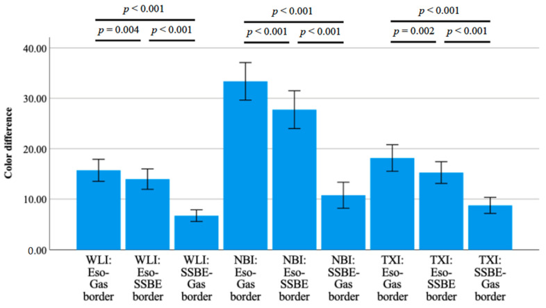
Figure 3.
Color differences among short-segment Barrett’s esophagus, esophageal, and gastric mucosa with three kinds of detection methods using third-generation high-vision GIF-1200N. Color differences between SSBE and esophageal or gastric mucosa in all detection methods were significantly smaller (p < 0.001 and < 0.001). Abbreviations: NBI: narrow-band imaging, SSBE: short-segment Barrett’s esophagus, TXI: texture and color enhancement imaging, WLI: white-light imaging.

