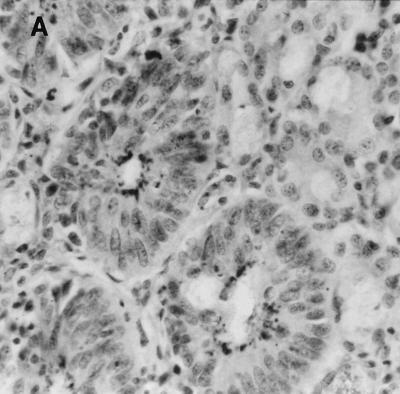
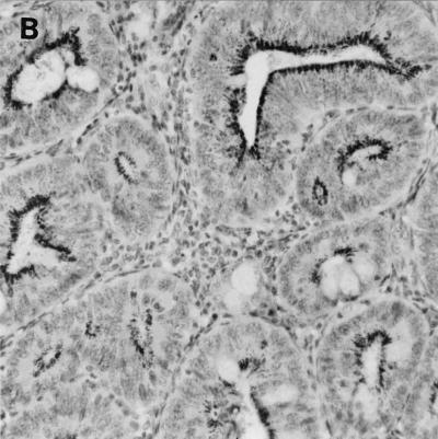
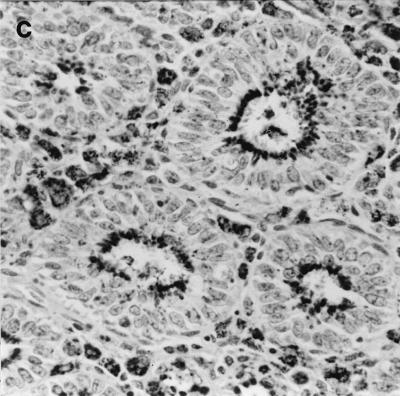
FIG. 3.
Immunochemistry of colonic tissue for L. intracellularis antigen, counterstained with hematoxylin. Dark-staining areas represent L. intracellularis organisms within epithelial cells. Infected crypts show epithelial hyperplasia. Original magnification, ×250. (A) Wild-type 129 mouse on day 28 post-challenge; (B) IFN-γR− mouse on day 28 postchallenge; C, IFN-γR− mouse that died (day 32 postchallenge).


