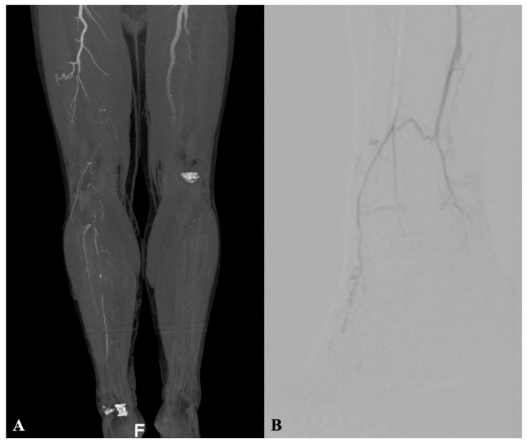
Figure 1.
(A) Pre-operative computed tomography angiography showing posterior view of right limb distal SFA, with popliteal and infrapopliteal tibial vessels occlusion without patent vessels crossing the ankle in the patient with Rutherford 4 and CS 4; (B) baseline angiography in patient with advanced forefoot gangrene, exhibiting distal posterior tibial occlusion, partial recanalization of distal anterior tibial, and peroneal arteries without target artery crossing ankle into foot; P2 patterns according to inframalleolar/pedal disease descriptor in the Global Limb Anatomic Staging System (GLASS).

