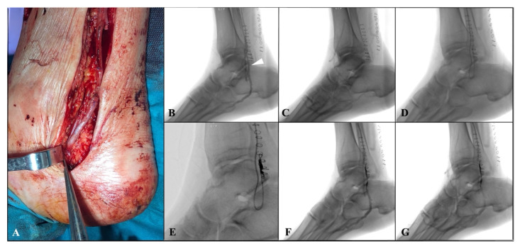
Figure 2.
(A) Intraoperative details showing the distal anastomosis performed at the level of the perimalleolar tibial vein in a termino-lateral fashion; (B,C) angiographic examination, performed 10 days after first surgery, revealing bypass patency and the main venous collateral, causing rapid wash-out of the contrast medium towards the leg, downstream of the distal anastomosis (white arrow); (D,E) selective catheterization of the collateral and embolization by a controlled-release spiral; (F,G) final angiography showing the occlusion of the main venous collateral and the implemented distal perfusion of the foot.

