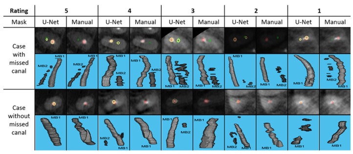
Figure 2.
Typical examples of each segmentation rating containing CBCT axial cross section with segmentation overlaid (top) and 3D view (bottom). MB1 is obturated canal, depicted in red and MB2 is unobturated canal, depicted in green. The yellow ring represents the location of manual segmentation relative to U-Net generated segmentation.

