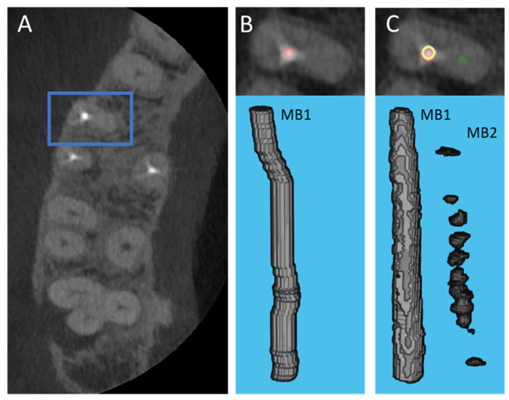
Figure 4.
Case where U-Net performed better than the manual segmentation; an unobturated MB2 canal was present, yet the resident had difficulty segmenting the canal. (A) CBCT axial cross section view showing the location of the root, blue box indicates the root; (B) Manually generated segmentation visualized in the CBCT axial cross section (top) and the corresponding 3D view (bottom), the red color depicts the superimposed obturated MB1 canal; (C) U-Net generated segmentation visualized in the CBCT axial cross section (top) and the corresponding 3D view (bottom), the U-Net generated canal segmentation is red for the obturated MB1 canal and green for the unobturated MB2 canal, the yellow ring represents the location of manual segmentation relative to U-Net generated segmentation.

