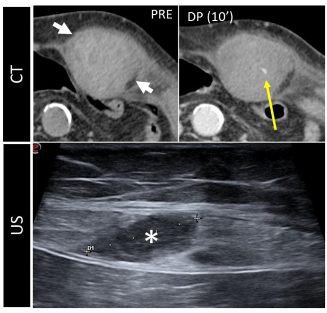
Figure 10.
CT and US post-traumatic rectus sheath hematoma above the arcuate line. CT scan shows a well-defined mass, hyperintense on basal scan, in the context of the left rectus sheath (short white arrows). In the second picture, the delayed phase acquired 10 min after the injection of intravenous contrast demonstrates active bleeding (yellow arrow). The third picture shows the same lesion (*), studied some days later on US.

