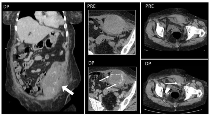
Figure 11.
Hematoma below the arcuate line in a 75-year-old woman, with acute myeloid leukemia and low platelet count. After a cough, she started complaining of strong abdominal pain and a mass started growing. Coronal and axial, pre- and post-contrast injection images showed the presence of a large left rectus sheath hematoma (thick arrow) with active arterial bleeding, as demonstrated by the subsequent spreading of contrast in all phases acquired (thin arrows). Since the hematoma was below the arcuate line, bleeding into the prevesical space is also noted (arrow, last picture, bottom row).

