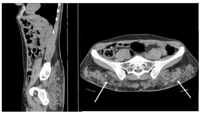
Figure 13.
Non-enhanced sagittal and axial CT scan demonstrates free silicon material scattered with an infiltrative appearance, expanding all along the subcutaneous fat of the posterior abdominal wall (arrows). Strand-like lesions coexist with nodular and plaque-like areas, making the differential diagnosis with neoplastic conditions difficult.

