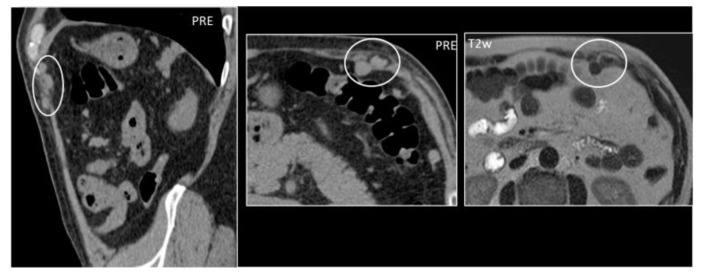
Figure 16.
A 59-year-old woman who underwent splenectomy some years prior. During a CT scan, new, rounded masses (circles) were found along the peritoneum and the left rectus abdominis muscle. These formations show parenchymal attenuation on CT (pictures 1 and 2) and share the same T2 intensity as the spleen. These lesions were later characterized as splenosis.

