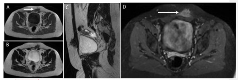
Figure 18.
MR study of an endometriotic nodule (arrows) in the left rectus abdominis muscles and subcutaneous tissue. Endometriotic implants inside muscle are well characterized on MR. They show heterogeneously low signal on T1 (A) and T2 images (images B,C) and strong enhancement after the injection of contrast agent (D). Sagittal reconstruction (C) shows the extension of the endometriotic nodule into fat tissue, fascia, and intramuscular location.

