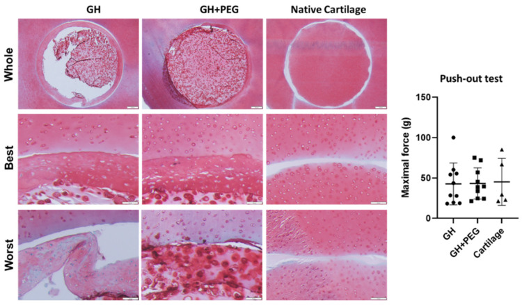
Figure 7.
In vitro repair experiment with bovine cartilage explant. H&E staining was used to assess tissue integration. The maximal force during the push-out test was recorded and compared among the three groups. Circles, squares, and triangles represent individual data points in GH, GH+PEG, and cartilage groups, respectively.

