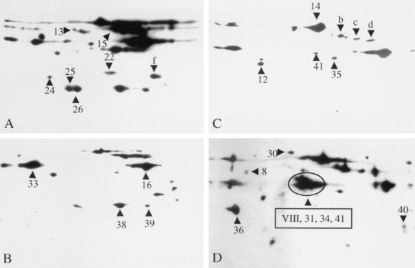
FIG. 3.
Two-dimensional Western blot analysis of stationary-phase GAS culture supernatant proteins with convalescent-phase sera obtained from mice with soft-tissue infection. Concentrated culture supernatant proteins (60 μg) were separated in the first dimension by isoelectric focusing (pH 3 to 10) and in the second dimension by SDS-PAGE with 12% (A and B) or 10% (C and D) gels. Concentrated culture supernatants from speB mutants of serotype M3 MGAS 315 (A and B) and serotype M1 MGAS 5005 (C and D) were used in the analysis. The separated proteins were transferred to nitrocellulose membranes, and Western blotting was performed with convalescent-phase sera obtained from mice infected with MGAS 315 (diluted 1:500) (A and C) and MGAS 5005 (diluted 1:1,000) (B and D). The numbered and lettered triangles indicate the proteins reactive in the Western blots that correspond to the protein spots (numbered identically) in Fig. 1.

