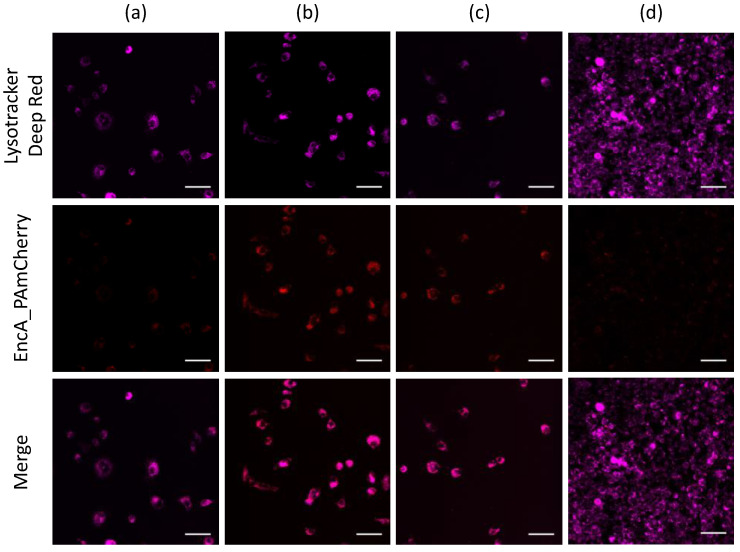
Figure 4.
Confocal imaging of RAW 264.7 cells after (a) 15 min, (b) 1 h, (c) 2 h and (d) 24 h of incubation with isolated EncA_PAmCherry. RAW 264.7 cells were imaged after irradiation with 405 nm laser for 30 s followed by 561 nm excitation of PAmCherry label (red fluorescence signal). Lysosomes were stained with LysoTracker Deep Red dye (purple fluorescence signal). Laser scanning confocal microscopy, Nikon Eclipse Ti2 and scale bars are 50 μm.

