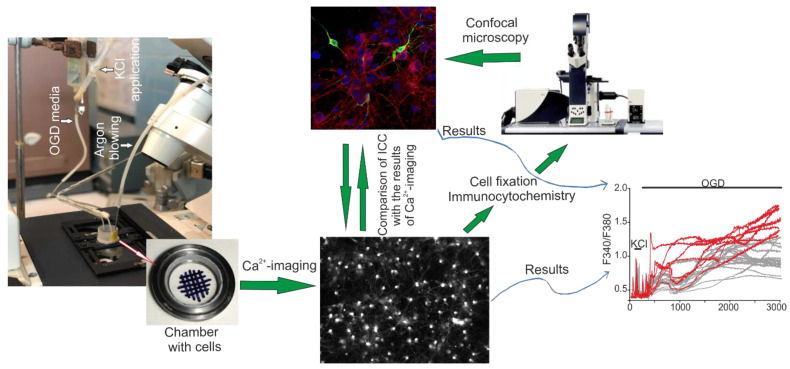
Figure 10.
Scheme of the original setup for creating OGD conditions for cells of the cerebral cortex in vitro and a method for comparing the results of Ca2+ imaging with the data of immunocytochemical staining of cell cultures. Cells of the cerebral cortex were grown on round coverslips and mounted in an experimental chamber, which was mounted on the object table of an inverted fluorescence microscope. A system for supplying OGD-media and blowing inert argon gas was connected to the chamber. After recording the [Ca2+]i dynamics, the cells were fixed and stained with specific antibodies. Next, the chamber with cells was transferred to an inverted confocal microscope, and using a grid, an area with cells was found in which the change in [Ca2+]i was recorded. The obtained confocal images were combined with a series of images obtained using Ca2+ imaging. Red curves is GABAergic neurons and gray curves is non-GABA-neurons.

