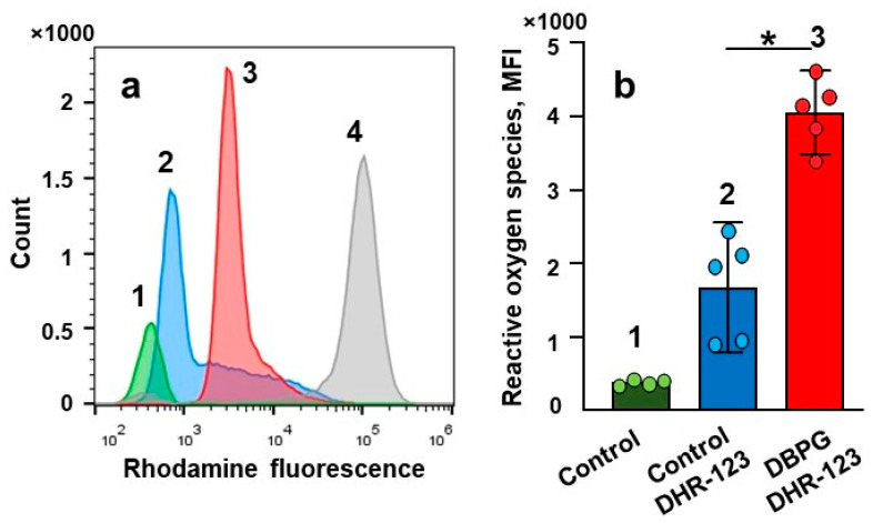
Figure 2.
Assessment of neutrophil activation in blood by flow cytometry. Whole blood was incubated at 36.7 °C for 1 h without any addition (1), and with the addition of DHR-123 and reagents. (2, 3, 4)—blood was incubated with DHR-123 for 15 min prior to addition of PBS (2, 20 µL to 250 µL of blood), DBPG (3, 2 mg of scaffold to 250 µL blood) and PMA (4, 150 nM). (a) Representative histograms of DHR-123 fluorescence in neutrophils. (b) Level of ROS produced in neutrophils after blood incubation without any additions (1, Control) and with DHR-123 and PBS (2, Control DHR-123) or DBPG (3, DBPG DHR-123). ROS production is reported as median fluorescence intensity (MFI). Data are means ± S.D., n = 5, * p < 0.05 versus Control-DHR123.

