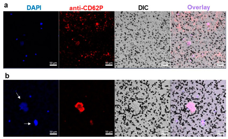
Figure 6.
Confocal microscopy of erythrocyte-poor blood samples incubated without addition (a) or with DBPG (b). DAPI—nuclear staining; anti-CD62P-PE—activated platelet staining. Black cells are erythrocytes. Arrows indicate accumulation of chromatin larger than 15 µm. DIC—differential interference contrast.

