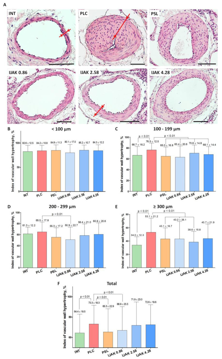
Figure 4.
Histological examination of lung vessels. (A) Representative micrographs of PA branches in studied groups (staining: hematoxylin–eosin, scale bar: 100 µm; red arrows indicate the thickness of the vascular wall). (B–F) Hypertrophy index of the vascular wall of PA branches: (B) vessels with outer diameter <100 µm; (C) vessels with outer diameter 100–199 µm; (D) vessels with outer diameter 200–299 µm; (E) vessels with outer diameter ≥ 300 µm; (F) all vessel diameters. INT—intact animals; PLC—placebo; PSL—prednisolone; IJAK—JAK inhibitor.

