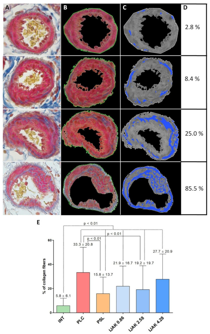
Figure 5.
The evaluation of the vascular wall fibrosis of PA branches. (A–C) Representative micrographs of vessels: (A) Picro-Mallory staining; (B) vessel border selection by Python plugin for fibrosis and IGH assessment 1.1 (ETU “LETI”, Saint Petersburg, Russia); (C) monochrome isolation of collagen fibers in the vascular wall structure; (D) nominal percentage of the vascular wall fibrosis of representative micrographs; (E) percentage of collagen fibers in the vascular wall structure of the PA branches in the studied groups. INT—intact animals; PLC—placebo; PSL—prednisolone; IJAK—JAK inhibitor.

