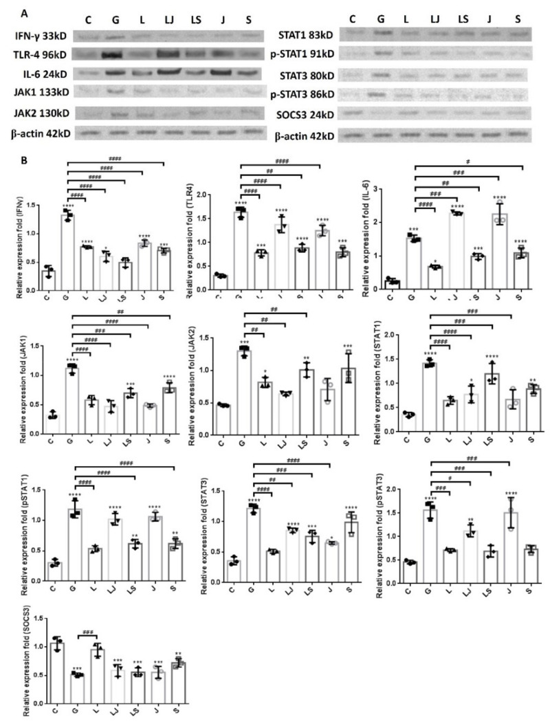
Figure 8.
JAK and STAT inhibitors affected the LCN2-downregulated JAK/STAT pathway and inflammatory factors in UPEC-infected bladder cells in a high-glucose environment. SV-HUC-1 cells were pretreated with a 25 μM JAK inhibitor (LJ) or 50 μM STAT inhibitor (LS) and 15 mM glucose for 24 h before a 25 μg/mL LCN2 pretreatment followed by UPEC infection (MOI of 100), as described above. Cells pretreated with the 25 μM JAK inhibitor (J) or STAT inhibitor (S) alone for 24 h followed by UPEC infection were used as respective controls. Cells pretreated with 25 μg/mL LCN2 alone (L) followed by UPEC infection were used as other controls. Cells infected with 15 mM glucose and UPEC alone were used as the respective positive controls (G). The picture shown is representative of a typical result. The total protein from all cell groups was collected for detection. (A) The total protein expression of JAK1, JAK2, STAT1, STAT3, phosphorylated-STAT1/STAT3, the inhibitor SOCS3, TLR4, IL-6, and IFN-γ were analyzed using Western blotting. (B) All data were normalized by their own internal reference, β-actin. The results were assessed using a densitometer and quantified using ImageJ software (NIH). The results are presented as means ± SDs of three independent experiments. * p < 0.05, ** p < 0.01, *** p < 0.001, **** p <0.0001 compared to the respective control groups. An analysis of variance was used to evaluate differences between various treatment groups and controls. Statistical differences between groups were determined using the Mann–Whitney U Student’s t-test. Statistical significance was set at p < 0.05. # p < 0.05, ## p < 0.01, ### p < 0.001, #### p <0.0001 for comparisons between the two individual groups.

