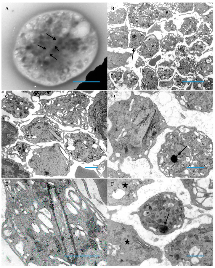
Figure 3.
Platelets from patient 2 demonstrate the character inclusions common for Medich syndrome. (A) This image demonstrates a faint outline (arrows) in a whole mounted, air-dried platelet; these are almost impossible to visualize when entirely within a cell, unlike inclusions that protrude from the cell and create a stark deformation of the discoid platelet shape (Original magnification 11,000×; Blue Bar = 1 micron). (B) Macrothrombocytes having vacuolated cytoplasm are readily apparent with one having a peripheral inclusion (arrow) (Original magnification 4800×; Blue Bar = 10 microns). (C) An inclusion in the center of this image is protruding from a platelet (Original magnification 8000×; Blue Bar = 1 micron). (D) Adjacent platelets demonstrate the two morphologic characteristics of Medich syndrome; one platelet has a giant alpha granule (arrow), whereas the second platelet has 2 scroll shaped inclusions (Original magnification 13,000×; Blue Bar = 1 micron). (E) The image demonstrates black granular aggregates of glycogen associated the multi-membranous “scroll” inclusion in a grey platelet (Original magnification 30,000×; Blue Bar = 1 micron). (F) In this image, two gray platelets (stars) absent of alpha granules are demonstrated and an additional platelet has a giant alpha granule (arrow) in contrast to the normal size of alpha granules populating the platelet (Original magnification 11,000×; Bar = 1 micron).

