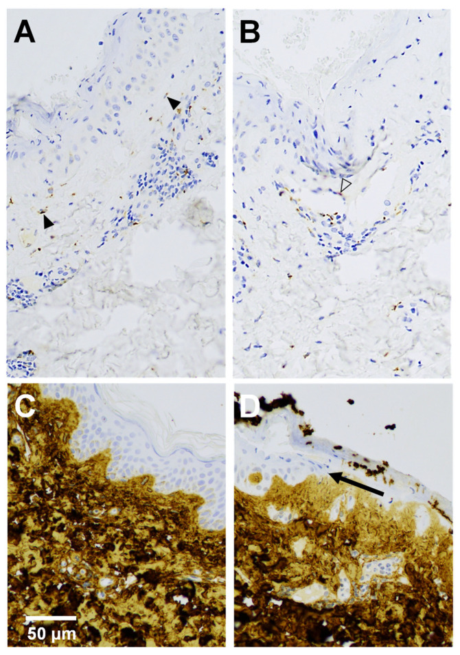
Figure 4.
TIMP-1 (A,B) and collagen α1 (I) (C,D) immunohistochemical analysis of a 4-day-old wound (A,B,D) with adjacent skin (C). (A) Fibroblasts (solid arrowheads) below neoepidermis and (B) endothelial cell (outline arrowhead) of small blood vessel show positive immunoreactivity of TIMP-1 in upper dermis. (D) Arrow indicates the tip of the migrating neoepidermal tongue.

