Figure 1.
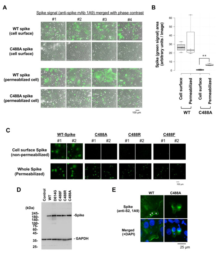
Loss of cell-surface expression of C488 mutant spike protein: (A) Wild-type (WT) and C488A mutant spike-expressing plasmids were transfected in Vero cells for 2 days. Cell surface spike protein was detected by anti-spike (1A9) antibodies and Alexa 488-conjugated secondary antibodies in non-permeabilized cells (upper panels). Intercellular spike protein was detected in Triton-X100 permeabilized cells (lower panels). (B) Green signals from spike-probed antibody in (A) were measured by ImageJ 1.44o. At least five images were analyzed in each setting. The spike-expressing area in C488A-expressing permeabilized cells was significantly larger than that in non-permeabilized cells. The Student’s t-test was performed to assess statistical significance. ** indicates p < 0.05. (C) Cell-surface expression of different C488 mutant spike proteins. WT and C488 mutant spike proteins were expressed in Vero cells for 2 days. Cell surface (upper panels) and whole intercellular (lower panels) spike proteins were detected, as in (A). The numbers #1 and #2 indicate two independent images. (D) Mutant spike protein expression was confirmed by Western blot analysis. GAPDH is shown as a loading control. (E) Subcellular localization of WT and C488A-mutant spike proteins in permeabilized Vero cells were analyzed by confocal microscopy. The green and blue signals represent spike proteins and DAPI-stained nuclei, respectively. The white arrowheads show spike proteins detected at plasma membrane.

