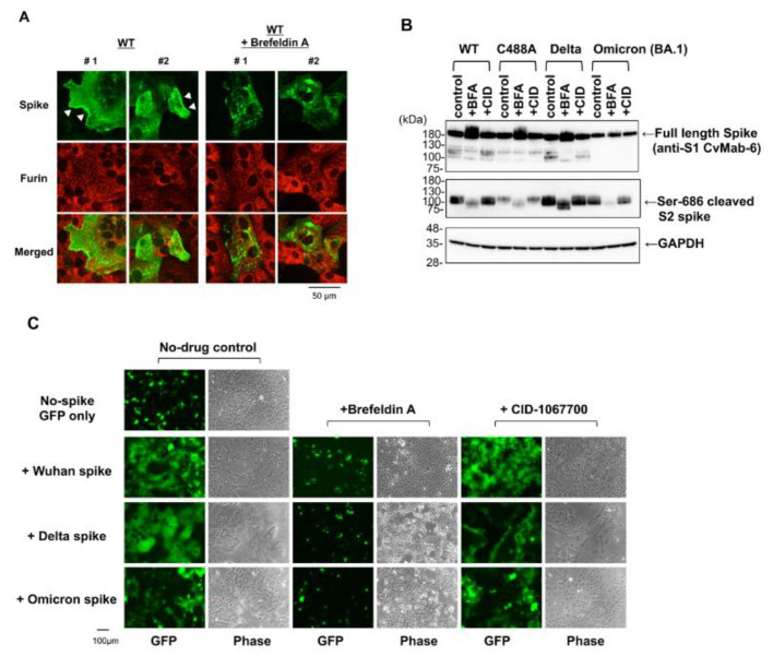
Figure 5.
Brefeldin A (BFA) interferes with SARS-CoV-2 spike protein function. (A) Vero cells were transfected with spike-expressing plasmid. The cells were treated with or without 1 µM BFA for an additional 15 h at 9 h post-transfection. The spike protein localization in permeabilized cells was examined using confocal microscopy. The numbers #1 and #2 indicate two independent images. White arrowheads indicate plasma membrane localization of spike protein. (B) HEK293T cells were transfected with spike-expressing plasmid. The cells were treated with 1 µM BFA and 1 µM CID-1067700 (CID) for additional 19 h at 9 h post-transfection. Full-length and Ser-686 cleaved spike proteins were detected by Western blotting with anti-spike (1A9) and cleaved SARS-CoV-2 spike (Ser686) antibodies, respectively. (C) VeroE6/TMPRSS2 cells were transfected with spike-expressing plasmid with pEGFP-C1. The cells were treated with BFA or CID-1067700 for an additional 15 h at 5 h post-transfection. The green fluorescence protein (GFP) signals and phase contrast cell morphologies were photographed with conventional fluorescence microscopy.

