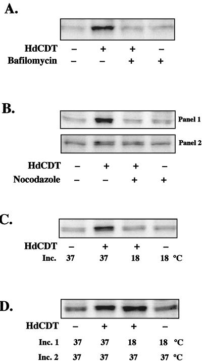
FIG. 5.
Effects of BafA1 (50 μM), nocodazole (30 μM), and 18°C incubation on tyrosine phosphorylation of cdc2 in HdCDT-treated cells. (A) Cells were exposed to BafA1 before and after toxin treatment. The postincubation time was 24 h. (B) Cells were exposed to nocodazole before and directly after toxin treatment (panel 1) or received nocodazole 60 min after toxin treatment (panel 2). The postincubation time was 8 h. (C and D) Cells were cultivated in eight individual petri dishes; four were kept as controls, and four were exposed to the toxin at the same time. (C) One pair of plates (HdCDT − and HdCDT +) was incubated (Inc.) at 37°C and the other was incubated at 18°C. Samples were prepared 12 h after toxin treatment. (D) One pair of plates was incubated at 37°C for 24 h. The other was incubated at 18°C for the first 12 h (Inc. 1) and then transferred to 37°C for another 12 h (Inc. 2). Blots are representative of three different experiments with each treatment.

