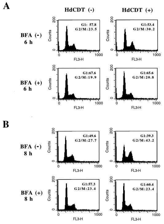
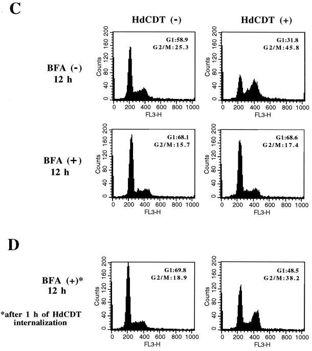
FIG. 6.
Effect of BFA on HdCDT-induced intoxication. Flow cytometry was used to analyze control [HdCDT (−)] and toxin-treated [HdCDT (+)] cells in the presence (+) or absence (−) of BFA. Cells were pretreated with BFA (2.5 μg/ml) for 45 min, exposed to the toxin, and postincubated in normal medium with BFA. Samples were prepared 6 h (A), 8 h (B), and 12 h (C) after toxin treatment. (D) Sample from cells treated with toxin, postincubated for 45 min at 37°C in fresh medium to allow internalization of the toxin, and exposed for 12 h to BFA. Percentages of cells in G1 and G2/M are shown. One representative experiment of three is shown. FL3-H, relative fluorescence.


