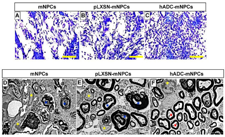
Figure 5.
Luxol fast blue staining of T9 spinal cord sections obtained from the mNPC (A), pLXSN-mNPC (B), and hADC-mNPC (C) transplantation groups at 6 weeks after SCI. The hADC-mNPC group (C) had smaller cystic cavities and more myelin sheaths than the other two groups (A,B). Scale bar = 20 μm. Transmission electron microscopy (TEM, 10,000×) of the lumbar section where mNPCs (D), pLXSN-mNPCs (E), and hADC-mNPCs (F) were transplanted after SCI. Transplanted hADC-mNPCs promoted axon remyelination at the lesion site. Red stars indicate remyelinated or mature axons in the hADC-mNPC group, while blue and yellow stars indicate degenerating myelinated axons and microglia/macrophage cells, respectively.

