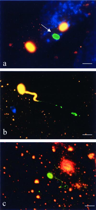
FIG. 1.
Differential immunofluorescence staining of MRC5 cells coincubated with E. cuniculi. The first labeling, using an anti-E. cuniculi antibody and a Cy3-conjugated secondary antibody (orange fluorescence), was performed without permeabilizing the host cells in order to label only extracellular spores. Host cells were then permeabilized with saponin followed by labeling with the same anti-E. cuniculi antibody visualized by a FITC-conjugated secondary antibody (green fluorescence). Extracellular microsporidia (spore walls and polar filaments) appear orange and intracellular ones appear green. Nuclei of MRC5 cells are stained blue (revealed by DAPI). (a) Intracellular, nongerminated spore (arrow). (b) Germinated extracellular spore inserting terminal portion of polar filament into host cell. (c) Germinated intracellular spore. Spore wall and entire polar filament are intracellular. Bar = 3 μm.

