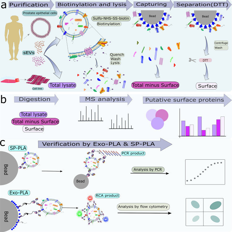
Fig. 1. Schematic illustration of the strategy to identify and validate sEV surface proteins.
a SEVs were isolated from human seminal fluids and PC3 culture media, respectively, and were treated with sulfo-NHS-SS-biotin to biotinylate outer membrane proteins on the surface of the sEVs before addition of lysis buffer. Biotinylated surface proteins were captured on streptavidin beads, and were released by DTT. b Total, total minus surface, and surface proteins were digested and identified by label-free semi-quantitative HRMS analysis. c The presence of these proteins was validated by Exo-PLA and SP-PLA.

