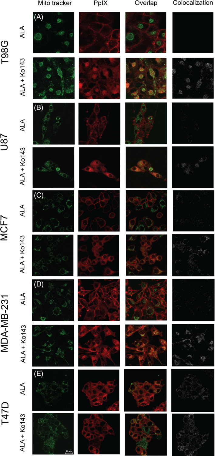
FIGURE 3.

Intracellular localization of the 5‐ALA‐derived photosensitizer PpIX for the various cell lines: A, T98G; B, U87; C, MCF7; D, MDA‐MB‐231; and E, T47D. The cells were incubated with 1.5 mM 5‐ALA for 4 hours with (odd rows) or without (even rows) pretreatment with Ko143 (1 μM). The cells were incubated with 100 nm MitoTracker Green 15 minutes prior to imaging. PpIX fluorescence is represented in red (left column) and MitoTracker fluorescence in green (second column). An overlay of PpIX fluorescence with MitoTracker fluorescence is shown in the overlap panel (third column) where colocalization can be seen in yellow. Pure colocalization between PpIX and MitoTracker is shown in the fourth panel in white
