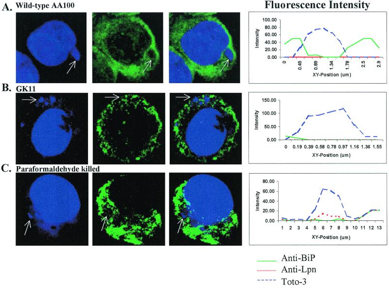
FIG. 6.
Colocalization of BiP with phagosomes of wild-type L. pneumophila (AA100), GK11, and paraformaldehyde-killed AA100 at 6 h postinfection. Nucleic acids were stained with Toto-3 (blue pseudocolor), which labels both intracellular and extracellular bacteria and the cell nucleus (first column); BiP was visualized using secondary antibodies conjugated to Oregon green (green pseudocolor; second column). Extracellular bacteria were visualized using secondary antibody conjugated to Alexa red (red pseudocolor). Colocalization is shown in the third column. Intensity of fluorescence across the indicated phagosomes (arrows) is shown at the right.

