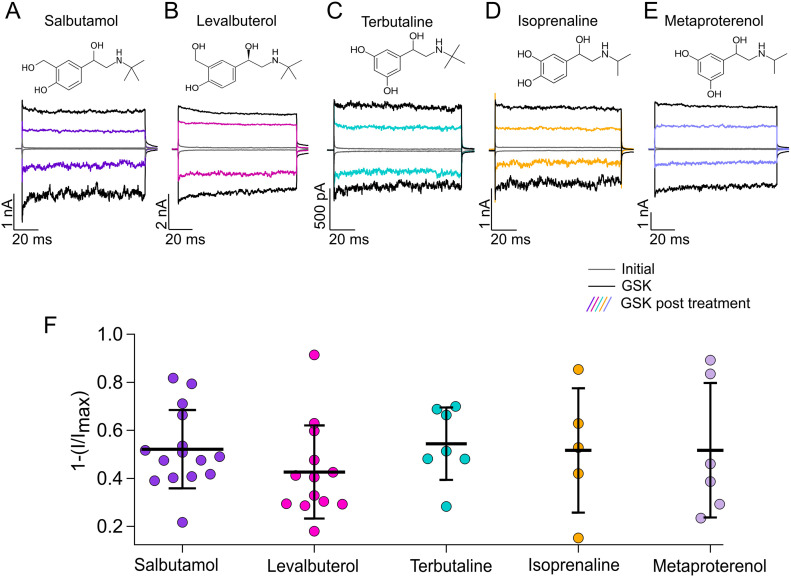
Figure 3. Effects of short-acting bronchodilator on TRPV4 currents.
(A, B, C, D, E) Representative traces of currents at +120 and −120 mV, obtained as in Fig 1A for different compounds. Trace colors represent gray for leak or initial currents, black for GSK 300 nM, and different colors for GSK after exposure of outside-out membrane patches to 500 µM of salbutamol, levalbuterol, terbutaline, isoprenaline, or metaproterenol. (F) Average data for experiments in (A, B, C, D, E). Data were normalized to the initial value with GSK. The percentages of inhibited currents after different treatments are as follows: 52.12% ± 16.3% after salbutamol (n = 15), 42.6% ± 19.4% after levalbuterol (n = 13), 54.4% ± 15% after terbutaline (n = 7), 51.6% ± 26% after isoprenaline (n = 5), and 51.7% ± 28% after metaproterenol treatment (n = 6). No statistically significant differences were found with one-way analysis of variance.
Source data are available for this figure.

