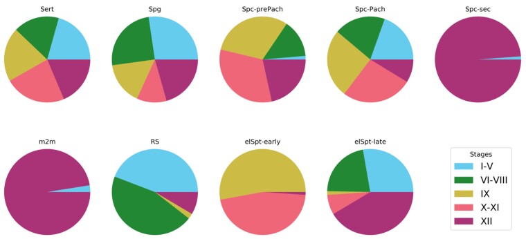
Figure 8.
Distribution of cell labels in different epithelial stages as extracted from STAGETOOL output. As expected, Sertoli cells were evenly distributed across the stages, whereas secondary spermatocytes (Spc-sec) and metaphase plates (m2m) were seen in stage XII, and occasionally in stage I to V (see Fig. 7C for explanation). Distribution of cell types in stage categories were accurate and biologically correct.

