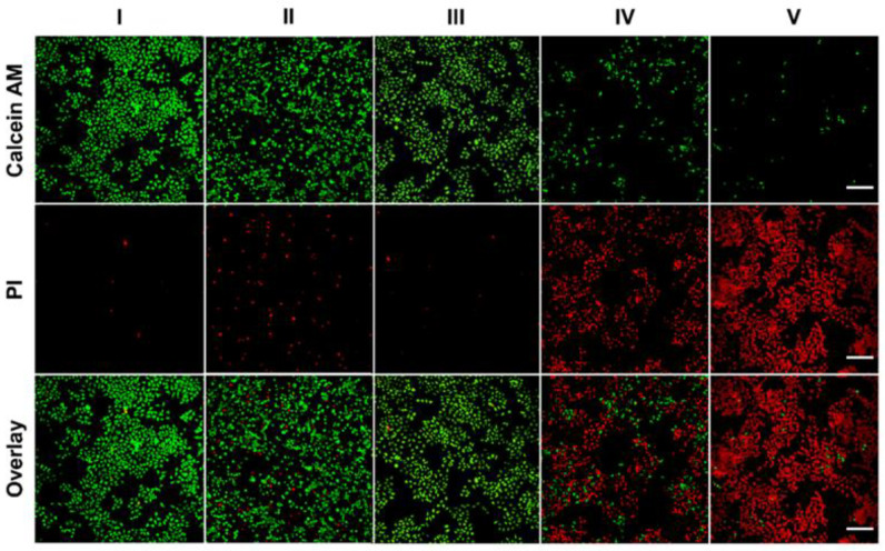
Figure 10.
The fluorescence inverted microscope was used to take fluorescent images of Hela cells stained with Calcium-AM (living cells) and PI (dead/late apoptotic cells) under different conditions. Scale: 200 µm. I. PBS, II. ICG—MTX, III. ICG—MTX@PPA Gels, IV. ICG—MTX with laser and V. ICG—MTX@PPA Gels with laser.

