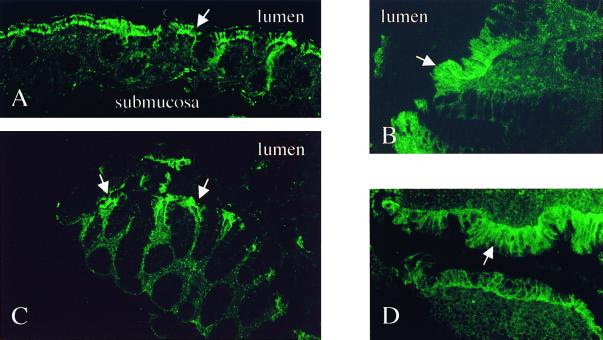
FIG. 6.
Constitutive TLR5 is predominantly expressed on surface IECs in healthy controls and remains unchanged in IBD. Shown are an overview (A) and detail (B) of normal, nondiseased mucosa of the colon; active, noninvolved CD colon cells (C); and active UC cells (D). White arrows indicate surface epithelial staining. Olympus AX70; original magnifications, ×40 (B and D) and ×10 (A and C); exposure times: 4 s (A), 3 s (B), and 5 s (C and D); Elitechrome Kodak Select series ISO400, Olympus exposure control unit PM-30, Superfluorescence 30 (exposure adjust setting, auto 2).

