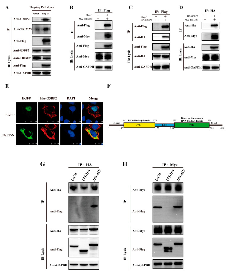
Figure 3.
Both the TRIM25 and G3BP2 protein are coimmunoprecipitated by the N protein. (A) The pcDNA3.1 vector and pcDNA3.1-N-Flag were transfected into cells. Twenty-four hours after transfection, TRIM25 and G3BP2 were precipitated by the N protein in pulldown experiments using the Flag tag. (B) The pcDNA3.1-TRIM25-Myc and pcDNA3.1-N-Flag were cotransfected into cells. Twenty-four hours after transfection, the interaction between the N protein and TRIM25 was detected by a coimmunoprecipitation experiment. (C) The pcDNA3.1-G3BP2-HA and pcDNA3.1-N-Flag were cotransfected into cells. Twenty-four hours after transfection, the interaction between N protein and G3BP2 was detected by coimmunoprecipitation. (D) pcDNA3.1-G3BP2-HA and pcDNA3.1-TRIM25-Myc were cotransfected into cells. Twenty-four hours after transfection, interaction between TRIM25 and G3BP2 was detected by coimmunoprecipitation. (E) Twenty-four hours after pcDNA3.1-G3BP2-HA and pcDNA3.1-N-Flag were cotransfected into cells, a cellular immunofluorescence assay was performed to assess the interaction between the N protein and G3BP2. Red fluorescence corresponds to the G3BP2-positive signal, green fluorescence labels SARS-CoV-2 N, and blue fluorescence corresponds to nuclei (DAPI staining; scale bar, 10 μm). (F) Diagram of the N protein divided into three parts: amino acids 1–174, 175–255 and 255–419. (G) pcDNA3.1-G3BP2-HA and pcDNA3.1-N (1–174/175–225/255–419)-Flag were cotransfected into cells. Twenty-four hours after transfection, an interaction between the 1–174 or 175–255 or 255–419 region of the N protein and G3BP2 was detected by coimmunoprecipitation. (H) pcDNA3.1-TRIM25-Myc and pcDNA3.1-N (1–174/175–225/255–419)-Flag were cotransfected into cells. Twenty-four hours after transfection, an interaction between the 1–174, 175–255 or 255–419 region of the N protein and G3BP2 was detected by coimmunoprecipitation.

