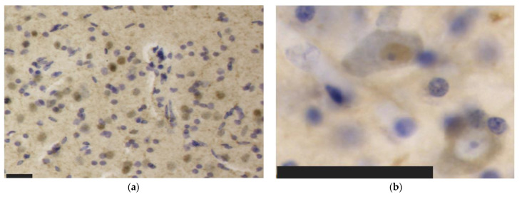
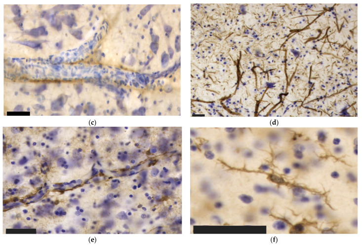
Figure 4.
Immunohistochemical patterns of the antibodies tested in this study in the bottlenose dolphin brain. Scale bar: 50 µm. (a) Aβ reactivity in neuronal nuclei and in the neuropil of the IC; (b) Nuclear (top left) versus cytoplasmic (bottom right) neuronal immunoreactivity to TDP-43 in IC neurons; (c) Perivascular immunoreactivity TDP-43 in the IC; (d) pNFP-immunolabeled thinner axonal fibers of the IC central nucleus alternating with much thicker external cortex axons; (e) Perivascular and intraparenchymal GFAP-expressing astrocytes in the IC; (f) Though the ramified morphology prevailed, few rod-shaped Iba-1-ir microglia were present in the VCN.


