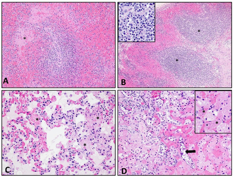
Figure 7.
Histological lesions following ASFV-MNG19 infection. (A) Spleen: Marked lympholysis and collapse of periarterial sheaths and fibrin thrombi (asterisk) in marginal zone (100×). (B) Renal lymph node: Marked lympholysis and necrosis (asterisk and insert) with loss of cortical and sinusoidal lymphocytes accompanied by marked filling and expansion of the subcapsular and medullary sinuses with hemorrhage and fibrin (100× and 400× insert). (C) Lung: Marked expansion of the alveolar spaces by congestion, fibrin thrombi (asterisk) and necrotic cellular debris, eosinophilic proteinaceous material; edema, fibrin and occasional macrophages fill the alveolar spaces (200×). (D) Kidney: Multifocal congestion and fibrin thrombosis (insert-asterisk) of the interstitial capillaries accompanied by hemorrhage and coagulative necrosis and dystrophic mineralization (arrow) of tubules (200×, 400×-insert).

