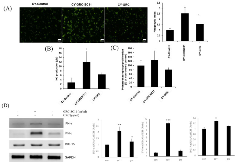
Figure 5.
Effects of GRC-SC11 on the phagocytic activity of peritoneal macrophages isolated from CY-treated immunocompromised mice. (A) Immunofluorescence images (left panel) of primary peritoneal macrophages from CY-treated immunocompromised mice orally administrated with distilled water, 1% GRC-SC11, and 1% GRC. Quantification of fluorescent beads up-taken by primary peritoneal macrophages from CY-treated immunocompromised mice (exposure for 48 h) (right panel). (B) Nitric oxide (NO) production of primary peritoneal macrophages isolated from CY-treated immunocompromised mice. Nitrite levels in the culture media were determined using Griess reagent and were presumed to reflect NO levels. (C) Cell proliferation of primary peritoneal macrophages was measured using a CCK-8 assay. (D) The levels of INF-γ, IFN-α, and ISG15 mRNA expression in RAW 264.7 cells. One-way analysis of variance was used to compare group means, followed by Dunnett’s t-test for the significance of individual comparisons (* p < 0.05, ** p < 0.01 and *** p < 0.001 vs. control group). Each figure is representative of three independent experiments: Scale bars = 50 µm.

