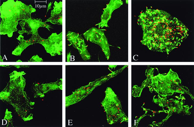
FIG. 7.
Confocal microscopy of bone marrow-derived macrophages infected (15 bacteria per cell) with LO28 (A to C) or the oppA mutant (D to F). Macrophages were observed at time zero (A to D), at 4 h (B and E), and at 8 h (C and F) postinfection. F-actin was stained with phalloidin (green). Bacteria were labeled with anti-Listeria antibodies (red). In the absence of OppA, there is a reduction of intracellular growth.

