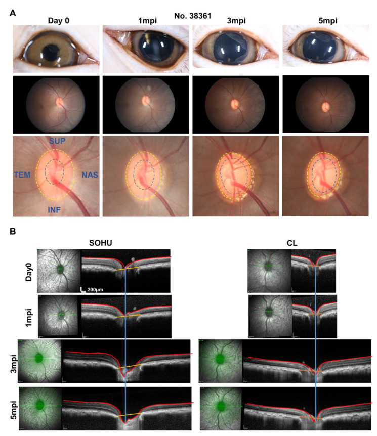
Figure 6.
ONH “cupping” in animal #38361 associated with IOP elevation. (A) The retinal fundus images of the SOHU eye before and after SO injection. Yellow dotted line outlines the optic disc; blue dotted line outlines optic cup; (B) Longitudinal SD-OCT imaging of macaque ON head with 48 radial B-scans acquired over a 30° area at 768 A-scans per B-scan, ART = 16 repetitions.

