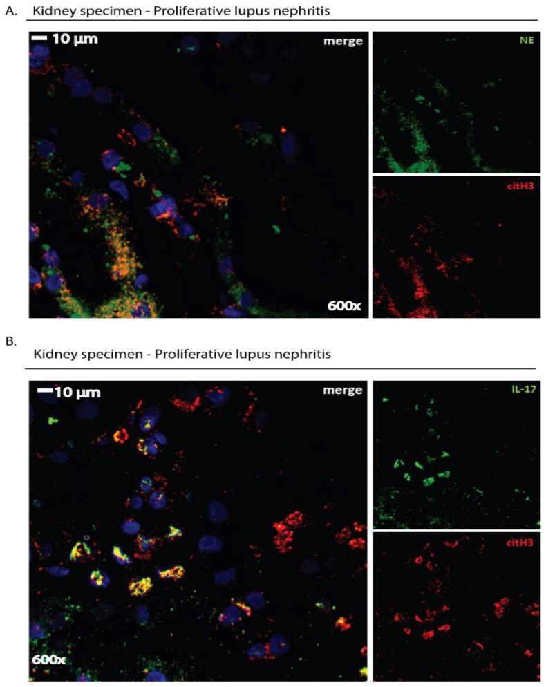
Figure 3.
Immunofluorescence staining: a qualitative tool to detect NET remnants or disease-related proteins located on NETs in human tissue specimens. Cross-sections (4 μm thickness) of kidney biopsy tissue derived from a patient with proliferative lupus nephritis (LN) were deparaffinized and stained with the appropriate antibodies. (A) NETs were visualized in kidney specimens as extracellular structures by staining with both a mouse anti-human neutrophil elastase ((NE), Abcam) antibody and a rabbit anti-human histone 3 (Anti-Histone H3 (citrulline R2 + R8 + R17), Abcam) antibody. (B) The presence of a disease-related protein on extracellular structures, such as IL-17 in LN, was examined using both a mouse anti-human IL-17A (R&D Systems) antibody and a rabbit anti-human histone H3 (Anti-Histone H3 (citrulline R2 + R8 + R17), Abcam) antibody. In (A,B), a goat anti-mouse AlexaFluor 488 antibody (Invitrogen) and a goat anti-rabbit AlexaFluor 594 antibody (Invitrogen) were used to detect primary antibodies. Sections were counterstained with DAPI and visualized in a confocal microscope (Revolution spinning disk confocal system; Andor, Belfast, UK). Data from the Molecular Hematology Laboratory archive are shown.

