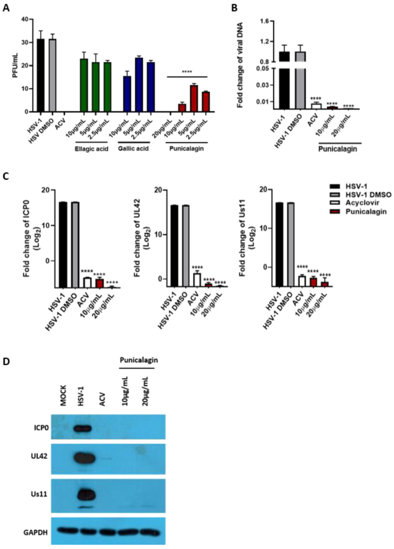
Figure 9.
Antiviral activity of pure compounds. (A) Vero cells and viral dilutions were incubated with punicalagin (20 µg/mL, 10 µg/mL, 5 µg/mL and 2.5 µg/mL), ellagic acid and gallic acid (10 µg/mL, 5 µg/mL and 2.5 µg/mL) for 1 h. The infection was then performed at 1 MOI with gentle shaking. After 1 h, the virus inoculum was removed and the monolayers were overlaid with a medium containing 0.8% methylcellulose in the presence of the tested compounds. Acyclovir (50 µM) was used as a positive control. The plaques were visualized after 3 days using crystal violet staining. (B,C) Relative quantification of viral DNA and viral transcripts (ICP0, UL42 and US11) was performed using real-time quantitative PCR and analyzed by the comparative Ct method (ΔΔCt). Values represent ± SD of the average of three independent experiments normalized against GAPDH. (D) Western blot analysis of ICP0, UL42 and Us11 viral proteins. GAPDH was used as a housekeeping gene. Data are expressed as a mean (± SD) of at least three experiments. **** p < 0.0001 vs. HSV-1 + DMSO.

