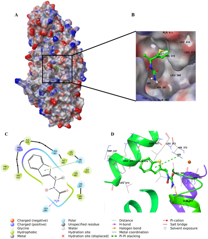
Figure 13.
Molecular docking of zileuton in stable human 5-LOX (PDB: 6N2W). (A) Molecular surface representation with solid style and electrostatic potential color scheme of red, white, and blue (min −0.3, max +0.3); (B) Zoomed image of zileuton in the active site of 5-LOX in molecular surface; (C) 2D representation of binding interactions of zileuton with amino acid residues in the active site within 4 Å distance; (D) 3D representation of zileuton in green color within the active site of 5-LOX. The H–bond and π–π staking interactions are in yellow and green dotted lines, respectively.

