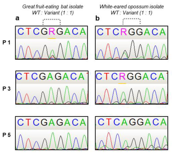
Figure 3.
Sanger sequencing chromatograms of a fragment of the G gene from the wild type and variants of RABV isolates during the competition assay. Dotted dashes highlight the mutated codon. Figure depicts passages 1, 3 and 5. (a) Great fruit-eating bat isolate (1069/18). (b) White-eared opossum isolate (2253/21). R represents a purine (adenine or guanine).

