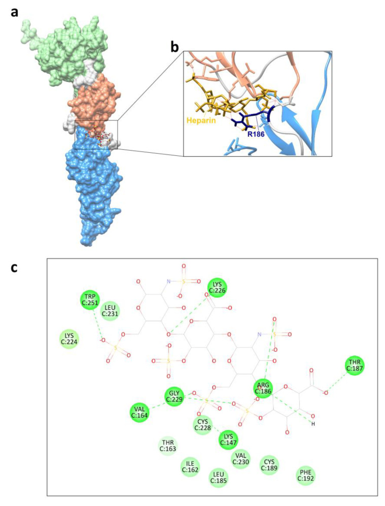
Figure 6.
Molecular docking results with heparin. (a) Three-dimensional model of mutant glycoprotein G186R+S188P with heparin. (b) Emphasis on the interaction of heparin (golden) and R186 (navy blue). (c) Two-dimensional diagram showing the glycoprotein amino acids involved in the hydrogen bond (darker green and dotted lines) and van der Waals interactions (lighter green) with heparin.

